by Monica Nenad, RDH, MEd, DHEd, CHES
October 28, 2024
This case has been prepared for publication during National Dental Hygiene Month 2024. The content is revealed as clues in email and on social media, with the full clues, case and care presented here. The case follows the ADHA Standards for Clinical Dental Hygiene Practice and incorporates a clinically supported solution from Colgate.
Patient Introduction & Assessment
The patient is a 55 year old Caucasian woman of Germanic ethnicity. She is physically fit, an avid tennis player, has never smoked, and takes no medications. The patient has had routine dental exams and prophylaxes throughout her life and reports never having dental decay or restorations. Her 3rd molars were removed in approximately 2014. The patient has a 10-year history of various basal cell and squamous cell carcinoma in situ located on the arm, shoulder, stomach, and leg, and in 2018, two melanomas located on the arm and leg. The patient sees a dermatologist every six months for full body skin checks.
In July 2022 the patient noticed a firm, pea-sized, nonmobile “bump” on her upper lip to the left of the philtrum. She reported the finding to her dermatologist that month at her routine appointment and also mentioned that she’d recently been hit in the face with a tennis ball in the same area. The dermatologist was not overly concerned, but due to the patient’s skin cancer history, referred her for an ultrasound of the area. The results came back as “consistent with bruising” and no treatment was provided.
At the patient’s next routine dermatology appointment in December 2022, she reported that she thought the bump had gotten larger and was more pronounced. The dermatologist referred her to an ophthalmic facial plastic surgeon for a biopsy. As the surgery progressed, the surgeon reported having to “chase” the affected tissue as the cancer had spread along the nerves and “burrowed” into an area midway up the nose and inner canthus of the eye. What was expected to take two hours became six.
First Diagnosis & Treatment
In January 2023 an intraoral biopsy was performed. The surgeon reported that the tumor tissue was “tough” and challenging to remove. The sample was sent to a lab and a diagnosis of “invasive basaloid carcinoma with infiltration” was returned. A surgery was immediately scheduled for February 2023 with a Mohs specialist. A two-hour procedure was anticipated and included excision of the tumor area under local anesthesia. The patient was treated with a Mohs approach and returned the next day for reconstruction. Tissue was taken intraorally from the bilateral buccal cheek pads and lower lip to fill in areas of extensive tissue removal. The patient healed uneventfully and after post-surgical follow-up appointments, treatment was considered complete.
Second Diagnosis & Treatment
In April 2023 the patient unexpectedly received a phone call from the treating surgeon. Additional biopsy samples obtained during the surgery had been sent to a specialist at a University teaching hospital for review. A new diagnosis of “mucoepidermoid carcinoma” which included melanoma was made. The patient was referred to a cancer treatment center for consultation with oncology specialists.
In May 2023 the patient was seen by a medical oncologist and radiation oncologist. The patient learned of the rarity of her tumor and despite a lack of definitive research regarding treatment, the recommended protocol for the new diagnosis differed from the treatment she had received. The patient was now advised to undergo six weeks of focused radiation five times per week for 12 minutes per session. Prior to initiation of radiation treatment, the oncologists referred the patient to a head and neck specialist to render an opinion regarding the surgical treatment that had already been provided and the therapy that was now planned. This physician had no additional recommendations.
The patient began radiation treatment in early June 2023. During all treatment sessions, the patient wore a radiation mask designed to position the head and direct the beam, in addition to a tongue displacement device intended to reduce radiation to that tissue. After 4 weeks she was advised to take a one week break due to side effects including extreme soft tissue sensitivity, open sores on the tongue, and severe xerostomia. After one week of healing time, the patient completed the final two weeks of treatment at the end of July 2023.
In consultation with her oncologists, the patient opted to forego chemotherapy following radiation treatment. A lack of research to validate use of chemotherapy to treat this type of tumor and the option of retaining it as a future treatment option should the need arise contributed to the decision-making process.
In late January 2024, six months after completing radiation treatment, the patient began routine follow-up care which included MRI evaluation with and without contrast every 6-8 weeks. Initial results demonstrated areas that were cautiously labeled “post-treatment inflammation”. Through four additional follow up appointments, these areas continued to show improvement lending support for the initial diagnosis. The patient is now on a 6-month evaluation protocol and due to be seen in December 2024.
Documentation
The patient remained conscientious of oral hygiene throughout diagnosis, treatment, and follow-up. Extreme sensitivity and discomfort of intraoral tissues during radiation treatment limited her ability to brush her teeth, but the patient reported that flosspiks were her “savior” as interproximal tissues were more tolerant. Although the left side of the patient’s face was treated, radiation “splatter” to the right side resulted in side effects throughout the oral cavity.
Following the first surgical procedure, the patient experienced significant tightening of tissues on the left side of her face as a result of the wide excision and suturing. To alleviate this condition and improve esthetic appearances, several reconstructive procedures were performed. In addition to cosmetic techniques used during the Mohs surgery, the patient was given Kenalog injections in an effort to “soften” scar tissue under the nose. Nine months after the initial surgery the patient underwent a procedure to create a left nasolabial fold using existing excess tissue. A subsequent procedure was performed in an attempt to raise the upper left lip and improve facial symmetry. The patient was informed that cosmetic fillers could be used in the lip area to further enhance appearance, but she has not opted for that procedure at this time. Throughout all post-surgical care, she was also advised to stretch her mouth and facial tissues as much and as often as possible and faithfully maintains this practice.
Following radiation treatment, the patient noticed an altered sense of taste; salty foods tend to burn and sweets are now preferred. The patient states that she is also aware of additional tightening of facial tissues. She noted she has lost “flexibility” of her lips and at times her “brain gets ahead of her mouth” resulting in a bit of stumbling when she speaks.
Although the patient was not advised to seek dental care prior to or during treatment, extreme xerostomia led her to seek help. The patient now uses a range of products to alleviate dryness and prevent tooth decay including salivary replacement sprays and rinses, PreviDent® 5000 toothpaste, and fluoride varnish.
At her most recent dental exam the patient was found to be decay free and put on a 6 month recall protocol. She was also told that hyperbaric oxygen therapy (HBOT) was a treatment option in the future if radiation damage becomes evident.
Follow-Up and Patient Outcomes
The patient’s prognosis is good. Her vigilance in monitoring her own health and awareness of her risk factors undoubtedly contributed to her early diagnosis and treatment of a rare cancer. She remains otherwise healthy and active and has resumed all life and social activities as before her diagnosis and treatment. The patient retains a positive outlook on her health and future.
____________________________________
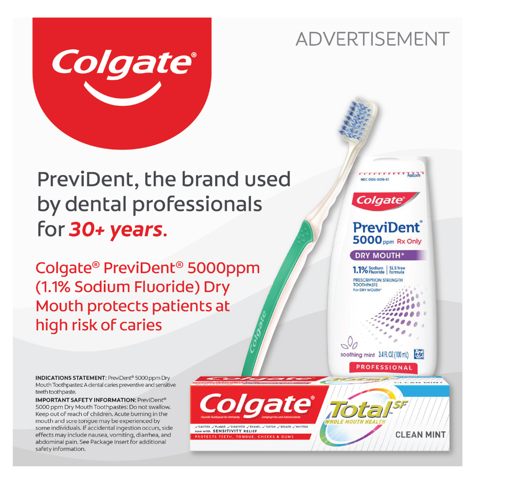 Patients of all ages can experience dry mouth, especially those who are medically compromised. Colgate® PreviDent® 5000 ppm Dry Mouth (1.1% Sodium Fluoride) toothpaste protects patients at high risk of caries.
To order contact your Colgate Territory Manager or call 1-800-2COLGATE (1-800-226-5428) Monday – Thursday (7:30 am – 5:30 pm CT) Friday (7:30 am – 4:30 pm CT) to speak to a customer service specialist.
References: 1. Joziak MT, et al. Comparison of enamel fluoride uptake and fluoride release from liquid and paste dentifrice. J Dent Res. 2003; 82 (Sp Issue). Abstract 1355. Important Safety Information: PreviDent® 5000 ppm Dry Mouth Toothpaste: Do not swallow. Keep out of reach of children. Acute burning in the mouth and sore tongue may be experienced by some individuals. If accidental ingestion occurs, side effects may include nausea, vomiting, diarrhea, and abdominal pain. See Package Insert for additional safety information.
STATUTORY PRICE DISCLOSURE FOR COLORADO PRESCRIBERS
In accordance with the Colorado Wholesale Acquisition Cost Disclosure Law (Colorado Revised Statutes Section 12-42.5-308), Colgate Oral Pharmaceuticals, Inc. is disclosing to you, as an authorized prescriber of prescription drugs in Colorado, certain required pricing information for Colgate Oral Pharmaceuticals, Inc. marketed prescription drugs. Please visit this webpage to access this price disclosure information.
STATUTORY PRICE DISCLOSURE FOR CONNECTICUT PRESCRIBERS
In accordance with the Connecticut Public Act No. 23-171, Colgate Oral Pharmaceuticals, Inc. is disclosing to you, as an authorized prescriber of prescription drugs in Connecticut, certain required pricing information for Colgate Oral Pharmaceuticals, Inc. marketed prescription drugs. Please visit this webpage to access this price disclosure information.
____________________________________
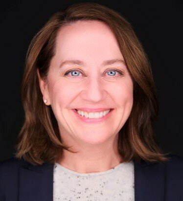 Dr. Monica Williamson Nenad is Faculty Chair/Program Director for Dental Programs at Rio Salado College in Tempe, AZ. She earned a BS in Dental Hygiene, holds a Masters in Education, and a doctoral degree in Health Education. Dr. Nenad is an ADHA Professional Fellow, a Certified Health Education Specialist (CHES) and completed a Graduate Certificate in Survey Research. She practiced clinical dental hygiene in a variety of specialties for the first half of her career with the second half being dedicated to dental and allied dental education and administration. Dr. Nenad is the current dental hygiene Commissioner on the Commission on Dental Accreditation (CODA).
